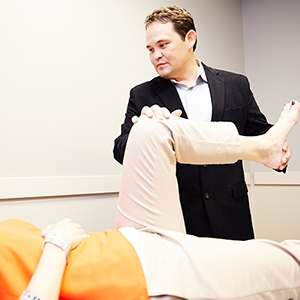
In March of 2016 we annouced Dr. Chin's involvement in a publication in Journal of Hip Preservation Surgery. Below is an update from Dr. Chin on his observances at SROSM in treating hip injuries.

During the past year, I have seen an increasing number of hip injuries present to our clinics. As our awareness and understanding of the pathology of these hip injuries increases, we are able to treat these hip injuries more effectively and efficiently, thus returning patients to their normal activity quickly. In addition, we strive to continue educating providers and patients about these injuries, so we can help more patients.
The field of hip arthroscopy is fairly new to the orthopedic armamentarium. We have made drastic improvements in our techniques and our instrumentation in the last 10 to 15 years which allows us to help patients who previously we were unable to offer any hope. As the awareness of hip injuries improves, we will be able to offer this groundbreaking treatment to more patients. Many injuries including athletic hip injuries, hip labral tears, hip cartilage injuries, hip ligament tears and laxity, hip strains, traumatic hip injuries, hip avascular necrosis, and hip arthritis can now be effectively treated using hip arthroscopy.
In general, hip arthroscopy is performed as a day surgery procedure. Patients undergo general anesthesia, and the surgery duration is approximately 1.5 to 3 hours. During the surgery, traction is placed on the operative leg in order to increase the size of the joint space to allow instruments to be introduced into the hip joint. Patients are well protected from traction related injury with special padding. Effort is made during the surgery to reduce the total time of traction in order to reduce the risk of injury. Most procedures are performed with less than one hour of traction time which is well within the safe zone for nerves. In addition, we can intra-operatively monitor nerves in order to improve the safety of the procedure. An arthroscopic camera is introduced into the hip joint and instruments are also introduced in order to repair damage to the labrum and cartilage, remove bone spurs and bony abnormalities, repair torn and stretched ligaments, repair torn tendons, remove loose bodies, and remove inflammatory bursitis.
Patients generally have a hip brace after surgery for 2 to 3 weeks and use crutches with partial weight-bearing for approximately four weeks. We use standard pain medications, but also use medications to inhibit scar formation and inhibit damaging enzymes. Patients begin immediate physical therapy in order to start range of motion of the hip which promotes healing and reduces scar formation. Patients are encouraged to use an exercise bike as early as one day after surgery and are instructed on passive and active range of motion exercises. Sutures are generally removed approximately two weeks after surgery and full weight-bearing can begin approximately four weeks after surgery. Most patients can expect to return to full activity and return to sports 3 to 6 months after their surgery depending on their injury and what procedures may have been performed.
Paul C. Chin, MD, PhD
Arthroscopy, Trauma, and Reconstructive Surgeon


