Conditions
-
Shin Splints

Shin splints are pain and inflammation of the tendons, muscles and bone tissue along the...
Know More -
Knee Injury
Pain, swelling, and stiffness are the common symptoms of any damage or injury to the knee.
Know More -
Unstable Knee
The knee joint is one of the largest joints in the body. This highly complex joint has several...
Know More -
Knee Sprain
Knee sprain is a common injury that occurs from overstretching of the ligaments that...
Know More -
ACL Tears
The anterior cruciate ligament (ACL) is one of the major ligaments of the knee.
Know More -
MCL Tears
The medial collateral ligament (MCL) is the ligament located on the inner part of the knee...
Know More -
MCL Sprains
Your MCL may get sprained or injured while twisting, bending or quickly changing direction.
Know More -
Meniscal Injuries
Meniscal tears are one of the most common injuries to the knee joint.
Know More -
Meniscal Tears
There are two wedge-shaped cartilage pieces present between the thighbone and the shinbone...
Know More -
Multiligament Instability
The knee is a complex joint of the body that is vital for movement. The four major ligaments...
Know More -
Knee Arthritis
The joint surface is covered by a smooth articular surface that allows pain-free movement...
Know More -
Knee Pain
Knee pain is a common condition affecting individuals of various age groups.
Know More -
PCL Injuries
Posterior cruciate ligament (PCL), one of the four major ligaments of the knee...
Know More -
Patellar Instability
Any damage to the supporting ligaments may cause the patella to slip out of the groove either partially...
Know More -
Patellar Tendon Rupture
The patellar tendon works together with the quadriceps muscle and the quadriceps tendon to...
Know More -
Posterolateral Instability
Posterolateral instability, also known as posterolateral rotatory instability (PLRI), is a...
Know More -
Knee Angular Deformities
Angular deformities of the knee are variations in the normal growth pattern during early...
Know More -
Knee Dislocation
Knee dislocation is a condition that occurs when the bones that form the knee joint...
Know More -
Distal Femur Fracture
The femur or thigh bone is the longest and strongest bone in the body, connecting the hip...
Know More -
Knock Knees
Knock knees, also called genu valgum, is a type of angular knee deformity in which...
Know More -
Terrible Triad Injuries
Terrible triad injury, also known as Unhappy triad or O’Donoghue triad, is a condition involving...
Know More -
Bowed Legs
Bowed leg is a bony deformity resulting in outward curvature of one or both knees of...
Know More -
Tibial Shaft Fracture
A tibial shaft fracture is a crack or break in the middle section of the tibia bone due to...
Know More -
Knee Fracture
A fracture is a condition in which there is a break in the continuity of the bone.
Know More -
Knee Sports Injuries
Trauma is any injury caused during physical activity, motor vehicle accidents, electric shock...
Know More -
Tibial Plateau Fracture
A tibial plateau fracture is a crack or break on the top surface of the tibia or shinbone in...
Know More -
Medial Meniscus Syndrome
Of the menisci within the knee, it is the medial that is more easily injured...
Know More -
Knee Stress Fractures
Stress fractures of the patella or knee are very rare. Approximately two out of 10,000 athletes...
Know More -
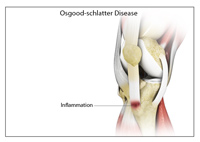
Osgood Schlatter Disease
Osgood-Schlatter disease is a common knee problem seen in growing adolescents.
Know More -
Multiligament Knee Injuries
Injury to more than one knee ligament is called a multiligament knee injury and may occur...
Know More -
Tibial Eminence Fractures
The tibial eminence, also called the tibial spine, is a bony protuberance of the tibia...
Know More -
Anterior Knee Pain
Anterior knee pain is characterized by chronic pain over the front and center of the...
Know More